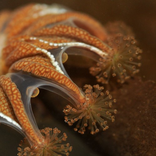
For quite some time, our group has been experimenting with using Vacuum sampling to get small, cryptic parts of the fauna off of coral reef rubble. The most recent, and successful, design is based on a sampler that Dr. Richard Pyle designed for his deep water work.
http://hbs.bishopmuseum.org/staff/pylerichard.html
We've had some trouble gettting the required parts through airline security, as Gustav has experienced first hand... but now it is all here and working! In the video below, Jenna and Mandy demonstrate its set up and use.
The device, though strange looking, has proven effective at getting at some spectacular animals that we have not previously collected using other methods, such as the snail below, a "witches' hat" from a family of snails that are parasitic on echinoderms (Eulimidae, Bacula morisyuichiroi).
-Sea
A blog about the field and lab adventures of the Invertebrate Zoology division at the Florida Museum of Natural History.
Sunday, November 14, 2010
Saturday, November 6, 2010
Lab Work
I know what you're thinking, "did you really go to a tropical paradise to toil in the hot sun and crystal water, and labor up idyllic forested slopes? Don't you ever take a break for some refreshing lab work, replete with the soothing glare of fluorescent lights, the gentle clink of scientific glassware, and the warm radiance of sterile surfaces?" Of course we do, except for sterile part. We're field biologists, the chemistry is upstairs.
I've already mentioned some of the work we do in the lab for processing specimens. First comes the sorting. Since this is year three of the project we really want to focus on getting things that are new and interesting, so people pick through the mass samples (sand sample, leaf litter, etc.) and pull out the animals, which are handed off to an expert to determine what qualifies . Usually one uses a microscope for this, but some larger stuff can be seen with the naked eye. Allow John to demonstrate.
Even an expert IDer needs help every once in a while. For this purpose we have photo field guides that we have compiled over the past two years, comprised of photos of specimens we have already collected. This also help ensure that we are collecting new animals, and not something we have collected many times in the past.
You can see one of the guides open next to Charlotte's microscope. That might be another one in her lap. The experts can also help by IDing animals in the guide whose identity has remained a mystery to us.
The specimens are also photographed, especially those that will be subsampled and will no longer be intact. We have several set-ups for this, including two microscope systems, but the basic system that we used for marine animals is a clear aquarium we built elevated off a black background.
After photography most of the animals are subsampled for DNA extraction. For this, a little snippet of tissue is placed in a 96 well plate which goes into the DNA-extracting-robot. Here, Jenna and Gary are about to start loading the plate in Jenna's hands (Jenna not pictured).
There's also a lot of data entry, as all the information that goes with each specimen (photo, subsample, location, identification, habitat, etc.) must be recorded for the Biocode database as well as for cataloging back at the museum. The lab work is not glamorous, but when we discover something new and exciting it often happens here. Please enjoy this reenactment.
Now that our numbers have swelled to 12, I'll try and keep up with the posting. Science waits for no one!
:) Mandy
I've already mentioned some of the work we do in the lab for processing specimens. First comes the sorting. Since this is year three of the project we really want to focus on getting things that are new and interesting, so people pick through the mass samples (sand sample, leaf litter, etc.) and pull out the animals, which are handed off to an expert to determine what qualifies . Usually one uses a microscope for this, but some larger stuff can be seen with the naked eye. Allow John to demonstrate.
The specimens are also photographed, especially those that will be subsampled and will no longer be intact. We have several set-ups for this, including two microscope systems, but the basic system that we used for marine animals is a clear aquarium we built elevated off a black background.
There's also a lot of data entry, as all the information that goes with each specimen (photo, subsample, location, identification, habitat, etc.) must be recorded for the Biocode database as well as for cataloging back at the museum. The lab work is not glamorous, but when we discover something new and exciting it often happens here. Please enjoy this reenactment.
:) Mandy
Monday, November 1, 2010
Underwater collecting techniques
Each collecting trip we try and take advantage of our numbers by employing a diverse array of sampling techniques. Everyone has their favored technique, usually dictated by the type of animal they are interested in, and the method most likely to yield that animal. I've been doing a lot of rubble brushing which entails picking a suitable rock from the bottom and brushing off the underside into a net. When I get out of the water at the end I empty the net into a plastic bag with water and bring it back to the lab for sorting.
This is me getting a nice, small rock from the bottom. Since I was manning this operation solo, I had three things to hold: the rock, the brush, and the net. A quick calculation reveals that that my two hands are somewhat inadequate. When the water is shallower this doesn't present a problem as I can kneel or stand (for breathing) and prop the rock (for brushing). At this depth I have to bring the rock to the surface to maneuver it (you know, to breathe), but it's hard to stay afloat while brushing and holding a boulder.
Art is a pro with the yabby pump. This device uses suction to draw animals out of burrows. The plunger is pulled quickly and the contents are emptied into a sieve. The sand falls through and whatever animal that was inhabiting that burrow is left in the sieve. Pumping is more difficult in the water because you don't have a nice solid surface with ample gravity to brace against. In this picture Sea is manning the pump and Art is holding the sieve on the lookout for interesting animals.
Art's particular interest is the shrimp that make many of the burrows, but there are often many commensals that come up in the pump as well. Like this cool gastropod (snail) which we're calling the Starship Enterprise, but which some call Phenacolepas.
We've also been doing a lot of sand sieving in our quest for micromolluscs. Since John is one of the resident mollusc guys he's being doing a lot of the sieving. He swims along the bottom pushing the sieve and scraping off the first few centimeters of sand. Then he roots through the sieve to see what he's found. As with the brushing, the contents of the sieve will be emptied into a plastic bag at the end of the dive.
The duration of each sieve push is limited by depth and buoyancy, and once again, when snorkeling, deeper is harder.
We've also been getting a lot of rubble samples now that Charlotte is here with her penchant for polychaet worms. This entails hauling around bags of rocks and coral rubble which will be rooted through back at the lab.
In gathering rocks and rubble there's a lot of sticking of hands into nooks and crannies, but one must be vigilant.
Can you see the eel? It's a dangerous job, but we're just the team to undertake it!
:) Mandy
Art is a pro with the yabby pump. This device uses suction to draw animals out of burrows. The plunger is pulled quickly and the contents are emptied into a sieve. The sand falls through and whatever animal that was inhabiting that burrow is left in the sieve. Pumping is more difficult in the water because you don't have a nice solid surface with ample gravity to brace against. In this picture Sea is manning the pump and Art is holding the sieve on the lookout for interesting animals.
We've also been doing a lot of sand sieving in our quest for micromolluscs. Since John is one of the resident mollusc guys he's being doing a lot of the sieving. He swims along the bottom pushing the sieve and scraping off the first few centimeters of sand. Then he roots through the sieve to see what he's found. As with the brushing, the contents of the sieve will be emptied into a plastic bag at the end of the dive.We've also been getting a lot of rubble samples now that Charlotte is here with her penchant for polychaet worms. This entails hauling around bags of rocks and coral rubble which will be rooted through back at the lab.
In gathering rocks and rubble there's a lot of sticking of hands into nooks and crannies, but one must be vigilant.
:) Mandy
Saturday, October 30, 2010
Waterfall Hike
We've had a couple new arrivals, and starting today our numbers are going to start increasing by leaps and bounds. This means a lot more trips to the field, with lots of different sampling techniques going on at once. And that means a lot more time in the lab for processing: anethetizing, photographing, subsampling, preserving. It's going to start getting crowded in here.
For one of our trips, Vetea (who lives and works here) took John, Yasunori, and I to look for terrestrial and freshwater molluscs in the area around Afareaitu Waterfall and the stream it feeds. The foliage was very dense in places, and we hiked around for about 4 hours fighting our way uphill through the vegetation.
But we did eventually make it to the waterfall.
Where we did some collecting around the pool at the base. Yasunori has a special interest in the nerite group of freshwater snails. He even found one that he had described as a species eight years ago.
While we were poking around the waterfall several more groups came and went, which was surprising considering how much of an effort it took us to get there. Then we realized there was a trail, which we weren't too proud to take on the way back to the car. Taking the trail down did nothing to diminish my sense of smug superiority over those who had taken the trail both ways!
So far every collecting trip has yielded new species for the biocode project, and my collecting skills are improving daily. Things are starting to get busy!
:) Mandy
So far every collecting trip has yielded new species for the biocode project, and my collecting skills are improving daily. Things are starting to get busy!
:) Mandy
Wednesday, October 27, 2010
My first Moorea snorkel
So on our second day we decided to get wet. Our focus was going to be micro organisms, mostly molluscs, so we were going to be doing brushing and sieving and sediment sampling in addition to general hand collecting. We headed out to a mud/silt flat with occasional coral bommies. Here is John looking for a suitable rock to flip and brush.
So, maybe I'll do some sand fanning, see if I can't find any...holy crap! Look at the size of that sea cucumber!
Wow, these guys are thick on the ground, I can see why Gustav likes it here. Ok, so, sand fanning. Maybe I can find some small snails or shrimp. Hey, is that an urchin!
It is an urchin! Those are pretty big too. They are crowded into every crevice in the coral. I can't believe how much coral is on this mud flat, and so close to shore. Looks like the fish like it too.
The colors are amazing! There are even picasso triggers (not pictured), my favorite! I wonder if this algae growth is hiding any...christmas tree worms!
I mean micro molluscs. I wonder if that algae is hiding any micro molluscs. Hmmm, so far nothing. Except a baby giant clam!
Is that a blue one!
This is pretty much how my snorkel went. I eventually began scouring the rocks along the jetty, and was excited to find several micro molluscs! Which turned out to be hermit crabs. So, there's a lot to see in Moorea and my first snorkel was full of new and exciting stuff. Next time I vow to do better. Maybe I'll help John with the brushing.
:) Mandy
:) Mandy
Tuesday, October 26, 2010
We're in Moorea!
This month begins the third and final installment of the three-year Moorea Biocode project. When our flight arrived yesterday at 5:30 am it was already full daylight. I can tell this will be something to get used to. The first wave to arrive was me, John, and Art. Art has been here before so he showed us the ropes. One of the first things we did was set up the lab (did you think I was going to say sleep, or eat, or shower...so did I).
Over the course of the next seven weeks we will be joined but twenty researchers coming and going as their time allows. We will be relying on their expertise to help us identify the animals in the groups that haven't been as thoroughly documented in years past. One of the holes in our knowledge is in phylum Mollusca, and one of the experts, Yasunori Kano, has already arrived to help out. So what does a group of scientists do after 13+ hours of travel, little to no sleep, inconsistently scheduled meals, and already having shown our dedication by setting up our stuff in the lab? We go collecting of course!
"Hmmm," you're thinking, "I thought you said you were in Moorea, you know, beaches, palm trees, coral reefs, all that jazz?" How right you are, but for our first collecting foray we decided to hit up some freshwater habitat which hasn't been as extensively sampled. John also found some interesting land snails in a tree on the edge of the stream, and Yasunori took a look around the estuary where the stream empties into the ocean and found some interesting animals as well. The estuarine area was full of these crabs:
But, we didn't collect any, they are so large and abundant that it's impossible that they were missed in the previous two years. After collecting we headed down to the grocery store to stock up on breakfast and lunch food. Sometime in the store I hit my wall and entered a quasi-zombie state. We put the food away and hit the lab for a while and then staggered off to our bungalows.
Only it was night. And now, please enjoy a picture of that jazz you were asking about earlier. We'll hit this up next.
:) Mandy
Monday, October 4, 2010
Invertebrates in the News will resume shortly meanwhile...
You can read about Triactis producta at the Invertebrate of the month. Andrea Crowther wrote about this sea anemone and its association with the pom-pom crabs Lybia.
Photo: Lybia tessellata, Réunion Island by François Michonneau/FLMNH CC 3.0
Sunday, October 3, 2010
Super Rewind: Keys Marine Lab 2010
I suppose I can't dive right into this post without acknowledging the extraordinary tardiness of it's presentation. Yes, I'm really lame, this is something we can all agree on. Now that that's out of the way, lets talk about our experience at the Keys Marine Lab (KML) in the Florida Keys after the Bioblitz in Biscayne Bay.
First off, the facility is great, there are dry labs, a wet lab, water tables (for keeping things alive), ample dorm space, and many other sciencey amenities including a boat complete with captain/dive master for diving or snorkeling. In the morning we would usually split into two groups one going out on the boat and the other doing shore-based collecting. Here are Gustav and Nat discussing strategy with the captain.
As far at the boat activities went, this strategy involved snorkeling, diving, and plankton tows. Because this was my first opportunity to use my AAUS Science Diver certification for actual collecting in the field I was pretty excited. I was also pretty inept. We'd all pile back into the boat after the air started running low, then while we were waiting for Gustav, who enters a presumably anaerobic state involving extremely low air consumption while diving, we would compare our finds of the dive. This Petrochirus diogenes was one of them.
I invariably lacked the collection skills of the more experienced among us (i.e. nearly everyone else) but I never came back completely empty-handed. And not only because my partner/dive buddy was the intrepid Sarah who had developed mad critter-spotting skills during her time in Moorea. Here she is on a snorkel excursion collecting some sargassum to be examined for critters.
The examining part looks like this.
Ok, maybe Hsiu is examining. The wet lab with its water tables and work space was where the animals were brought and sorted. This was also where sargassum was picked through and chunks of coral rubble were cracked open to reveal the creatures within. I think Nat is taking something from the water table back to the dry lab to be processed. The dry lab was a hive of activity. As earlier at Bioblitz, and as is usual in general in the field, the animals are anesthitized, photographed, and subsampled. There is also someone manning the computer and entering the animals into a spreadsheet along with the location they were found and their taxonomic ID. Here are Gustav and Anne DuPont consulting over an ID. Jenna is subsampling, and Francois is at the photo station. Since I don't see anyone next to Jenna where the computer person usually sat, I'm going to assume that it was me, who paused ever-so-briefly to take this picture.
After a long day of collecting and processing we'd haul our carcasses up to the dorm for dinner. Carole even made us some brownies to keep our energy and spirits high. Don't George, John, and Anne look revitalized?
In all, it was a very fun and productive trip. As we aim to increase our knowledge of the Florida fauna, we will hopefully be spending more time at KML in the future.
(Thanks to Carole Marshall and Anne DuPont for the crab picture and the brownie picture!)
First off, the facility is great, there are dry labs, a wet lab, water tables (for keeping things alive), ample dorm space, and many other sciencey amenities including a boat complete with captain/dive master for diving or snorkeling. In the morning we would usually split into two groups one going out on the boat and the other doing shore-based collecting. Here are Gustav and Nat discussing strategy with the captain.
I invariably lacked the collection skills of the more experienced among us (i.e. nearly everyone else) but I never came back completely empty-handed. And not only because my partner/dive buddy was the intrepid Sarah who had developed mad critter-spotting skills during her time in Moorea. Here she is on a snorkel excursion collecting some sargassum to be examined for critters.
In all, it was a very fun and productive trip. As we aim to increase our knowledge of the Florida fauna, we will hopefully be spending more time at KML in the future.(Thanks to Carole Marshall and Anne DuPont for the crab picture and the brownie picture!)
Sunday, June 20, 2010
Rewind: BioBlitz 2010
Back in April/May our lab participated in the National Geographic BioBlitz down in Biscayne Bay. BioBlitz is a 24 hour biodiversity survey (from noon to noon), where we document as many species as possible. Groups of scientists lead groups of science enthusiasts on short forays to see what we can find using various collecting methods. We were involved in both the marine and terrestrial invertebrate surveys. There was a base of operations at Convoy Point, but we were involved with the survey based out of Elliot Key. While the tour groups would come and go throughout both days we the scientists spent two action packed nights camping on Elliot Key. So what does it look like when 12 scientists go camping? Something like this.
I realize that's it hard to really grasp the scale of supply-mountain, but we and our gear filled the deck of our the boat that ferried us to the island. And it was all deck. It was a landing-craft designed for transporting vehicles.
The eating/sciencing portion of the excursion was conducted on one side of the island, and the sleeping portion was conducted on the other. To travel between them one had to go through The Gauntlet.
I know, it's so scenic and tranquil. We too were lulled into a false sense of security, but a few steps into The Guantlet and we realized why the woman on the boat had been so insistent about bug spray.
What appears to be a diseased limb swiftly traveling down the road to amputation is actually a buffet of culinary delights from the perspective of the aggressive and bountiful mosquito and no-see-um populations of the island.
So how did we avoid the bugs (besides employing hazardous level of DEET)? By getting in the water! In this picture Jenna and Sam are showing a school group how to use a yabby pump and sieve to collect critters from burrows in the sediment. I realize you can't see the yabby pump in this picture, but the good news is I'm not holding out for a career in photojournalism.
Unfortunately I also don't have any pictures of John braving the wooded areas searching for land snails. Except for my sprints through The Gauntlet necessitated by some desperate need (food, sleep, more bug spray), I avoided the woods whenever possible.
Throughout the day, groups would come in from collecting trips and some animals would be displayed in trays on outside tables so that people could admire them. A group of us would man the tables to tell people about what they were looking at. For instance, dig this lobster.
Now dig Jenna, Nat, Gustav, and François showing this group of children a thing or two about biodiversity.
That tent in the background was the headquarters for this operation. Many animals were admired and released, but many also ended up in the tent where they were anesthetized. Then they were photographed, subsampled, and preserved to go back with us to the museum. Sarah is photographing, while Julie and Hsiu are subsampling.
Having a physical specimen, accompanied by a live photo and DNA sequence, allow diversity information to be preserved in such a way that others can use it for their own research.
All in all it was a lot of work and a lot of fun, and we expanded our knowledge and collections of Florida's biodiversity, but we weren't done yet. After BioBlitzing most of us continued taking our show on the road and headed down to the Keys Marine Lab on Long Key. More on that in another post.
What appears to be a diseased limb swiftly traveling down the road to amputation is actually a buffet of culinary delights from the perspective of the aggressive and bountiful mosquito and no-see-um populations of the island.So how did we avoid the bugs (besides employing hazardous level of DEET)? By getting in the water! In this picture Jenna and Sam are showing a school group how to use a yabby pump and sieve to collect critters from burrows in the sediment. I realize you can't see the yabby pump in this picture, but the good news is I'm not holding out for a career in photojournalism.
Throughout the day, groups would come in from collecting trips and some animals would be displayed in trays on outside tables so that people could admire them. A group of us would man the tables to tell people about what they were looking at. For instance, dig this lobster.
All in all it was a lot of work and a lot of fun, and we expanded our knowledge and collections of Florida's biodiversity, but we weren't done yet. After BioBlitzing most of us continued taking our show on the road and headed down to the Keys Marine Lab on Long Key. More on that in another post.
Friday, June 11, 2010
Sea cucumber workshop in Guam
Part of the lab is currently in Guam because of sea cucumbers.
In the context of the PEET project, we brought together 15 sea cucumber specialists and students -- from 10 countries -- in Guam to share knowledge about field and lab techniques for taxonomic studies of our favorite echinoderm group. We are now half-way through the workshop (it started on June 7th) and we have been very busy.
Our days are filled with field trips where we collect sea cucumbers in their habitats, time in the lab where we dissect the animals collected and look at their ossicles and time in the classroom where sea cucumbers specialists give lectures and students present their research.
We have been really successful in the field and found several species that haven't been recorded from Guam before, and even maybe a couple of new species. There are two main reasons for these new records.
First, it highlights the progress that the PEET project has made since it started. By visiting museum collections to find type specimens, by collecting sea cucumbers in many places to understand morphological variation in species and by sequencing all this freshly collected material, we have a better idea of the characters that are useful to identify sea cucumber species and we have refined our understanding of some enigmatic species. For instance, there is a genus of sea cucumber called Labidodemas that live among coral rubble in lagoons. In the past, most Labidodemas were assigned to a species called semperianum. The reason is simple, most Labidodemas species look almost alike: they are whitish, have long tube feet on the ventral side and a dark red/brown coloration around the anterior and posterior ends. However, by looking at their ossicles (microscopic skeletal elements found in their skin), it turns out that it is fairly easy to identify the other species of this genus. During the workshop, we thus recorded for the first time in Guam the species called Labidodemas pertinax. By looking carefully at the live specimen, we are now able to tell the two species apart.
The image at the top is Labidodemas semperianum and the one at the bottom is Labidodemas pertinax. Can you see the difference in the color of the tentacles and the tube feet? (click on the images to enlarge)
The other reason that we are finding new records and new species in Guam, is because it's the first time there are so many people looking for sea cucumbers at the same time. It's amazing what 15 pairs of trained eyes can see when they are specifically looking for sea cucumbers.
I'm sure we'll catch many more interesting things before the end of the workshop and I'll keep you posted!
Monday, May 24, 2010
Invertebrates in the News #5 (follow up)
Last week, I told you that Terry Gosliner was blogging about his field trip in the Philippines where he is looking for nudibranchs (sea slugs).
Today, there is a new post about his adventures.
Update: Here are his final posts that wrap-up his expedition.
Today, there is a new post about his adventures.
Update: Here are his final posts that wrap-up his expedition.
Thursday, May 20, 2010
Invertebrates in the News #6 - DNA reveals the complex life cycle of the stalked jellyfishes
If I say jellyfish, you probably picture a Frisbee-like animal with long tentacles that swims gracefully in an open ocean.
However, there is a group of jellyfishes (the Stauromedusae) that do things a little differently. First, they don't swim but live attached to the bottom with a stalk, hence their common name: the stalked jellyfishes.
Second, they are thought to have a "simple" life cycle. Most jellyfishes
go through several stages before they reach adulthood. The typical
jellyfish starts life as a small polyp (something that looks like a sea anemone) attached to the ocean floor. This polyp buds small medusae (jellyfishes) that grow until they reach sexual maturity. At this point, they release gametes into the ocean. After fertilization, the egg forms a planula larva which will settle on the bottom to give rise to a new polyp.
In stalked jellyfishes, things were assumed to be a little different, but simple: the planula attaches to the bottom and metamorphoses into a polyp (so far everything is normal). The top of the polyp metamorphoses into something that looks like a jellyfish while the bottom part stays the same and still looks like a polyp; hence a stalked jellyfish. The only problem with this story is that it is based on only a few observations based on few species, and it's hard to know if all the Stauromedusae do the same thing.
There is another problem: how can you tell if the two stages of the life cycle that look very different belong to the same species? In the best case, you can observe the full life cycle in the field or in the lab, but it is not always possible to do so. In other cases, you can only observe part of the life cycle.
In 1996, Gerhard Jarms & Henry Tieman published a paper where they described a new species of hydrozoan polyps that live on clam shells of the Antarctic that they named Microhydrula limpsicola. They kept the animals in the lab for 4 years, and only observed asexual reproduction by frustules (larvae produced asexually).
In a recent paper, Lucilia Miranda and her colleagues demonstrated that Microhydrula limpsicola is just a stage in the life history of the stalked jellyfish Haliclystus antarcticus. They were able to connect these two life stages by discovering that the DNA of the polyps (what was thought to be M. limpsicola) and the stalked jellyfish (H. antarcticus) were identical.
The life cycle of the stalked jellyfishes is thus much more complicated than we thought.
Furthermore, Microhydrula limpsicola was not the only one of its kind. There are two other species that were thought to belong to this family (the Microhydrulidae). It will now be interesting to determine if they are also stages of other species of stalked jellyfishes.
Links
Credits
However, there is a group of jellyfishes (the Stauromedusae) that do things a little differently. First, they don't swim but live attached to the bottom with a stalk, hence their common name: the stalked jellyfishes.
Second, they are thought to have a "simple" life cycle. Most jellyfishes
go through several stages before they reach adulthood. The typical
jellyfish starts life as a small polyp (something that looks like a sea anemone) attached to the ocean floor. This polyp buds small medusae (jellyfishes) that grow until they reach sexual maturity. At this point, they release gametes into the ocean. After fertilization, the egg forms a planula larva which will settle on the bottom to give rise to a new polyp.
In stalked jellyfishes, things were assumed to be a little different, but simple: the planula attaches to the bottom and metamorphoses into a polyp (so far everything is normal). The top of the polyp metamorphoses into something that looks like a jellyfish while the bottom part stays the same and still looks like a polyp; hence a stalked jellyfish. The only problem with this story is that it is based on only a few observations based on few species, and it's hard to know if all the Stauromedusae do the same thing.
Stauromedusae (stalked jellyfish) - Haliclystus antarcticus
There is another problem: how can you tell if the two stages of the life cycle that look very different belong to the same species? In the best case, you can observe the full life cycle in the field or in the lab, but it is not always possible to do so. In other cases, you can only observe part of the life cycle.
In 1996, Gerhard Jarms & Henry Tieman published a paper where they described a new species of hydrozoan polyps that live on clam shells of the Antarctic that they named Microhydrula limpsicola. They kept the animals in the lab for 4 years, and only observed asexual reproduction by frustules (larvae produced asexually).In a recent paper, Lucilia Miranda and her colleagues demonstrated that Microhydrula limpsicola is just a stage in the life history of the stalked jellyfish Haliclystus antarcticus. They were able to connect these two life stages by discovering that the DNA of the polyps (what was thought to be M. limpsicola) and the stalked jellyfish (H. antarcticus) were identical.
The life cycle of the stalked jellyfishes is thus much more complicated than we thought.
Furthermore, Microhydrula limpsicola was not the only one of its kind. There are two other species that were thought to belong to this family (the Microhydrulidae). It will now be interesting to determine if they are also stages of other species of stalked jellyfishes.
Links
- Miranda LS, Collins AG, Marques AC (2010) Molecules Clarify a Cnidarian Life Cycle – The “Hydrozoan” Microhydrula limopsicola Is an Early Life Stage of the Staurozoan Haliclystus antarcticus. PLoS ONE 5(4): e10182. doi:10.1371/journal.pone.0010182
- Jarms G & Tiemann H (1996) On a new hydropolyp without tentacles, Microhydrula limopsicola n. sp., epibiotic on bivalve shells from the Antarctic. Sci. Mar. 60(1): 109-115 http://www.icm.csic.es/scimar/index.php/secId/6/IdArt/2742/
Credits
- Photo of jellyfish (top) François Michonneau. Lizard Island, February 2009. CC 3.0
- Photo of H. antarcticus and drawing of life cycle by Miranda et al 2010. CC 3.0
- Photo of the close up of a stauromedusa by Minette Layne CC 2.0
Wednesday, May 19, 2010
Invertebrates in the News #5
Currently, the New York Times is hosting a blog series entitled "Scientists at work, notes from the field".
Today, Terry Gosliner from the California Academy of Sciences writes about new species of nudibranchs he found in the Philippines.
You can read his blog posts here.
Today, Terry Gosliner from the California Academy of Sciences writes about new species of nudibranchs he found in the Philippines.
You can read his blog posts here.
Images by Terry Gosliner
Subscribe to:
Posts (Atom)