Sunday, October 30, 2011

ARMSed and Ready

Hey, remember all that time when I wasn't posting?  One of the things we were doing was survey dives in the Gulf off Cedar Key and Steinhatchee looking for suitable habitat to deploy some ARMS.  (You might remember ARMS from this post).  This is a continuation of the survey work we did on the R/V Weatherbird back in March (remember this or that or this other post), but the water was much warmer this time, approaching 90 degrees!  We were looking for hard-bottom areas, which turned out to be rarer than we might have anticipated.  Most of Gustav's quick peeks at the bottom revealed long stretches of sand, but a few times we found what we were looking for and went down to do a more extensive survey of the area.  These areas had lots of sponges and Caulerpa algae


and lots and lots of tunicates of various types


So now that we'd found some potential sites to set up some ARMS, where were we going to get them?  Thank goodness for Woody at the Smithsonian Research Station in Ft. Peirce.  They had some old disassembled ARMS that they were willing to let us use, and Woody even put them together for us (except for the base for ease of transport).  So what do 27 ARMS look like after they've been driven back to Gainesville and piled on a cart?
It sure would be foolish for one person to put 367 pounds of metal and pvc all together on a cart and then expect to move that cart over a non-flat surface...unless that person were a glutton for punishment, or placed well in an arm-wrestling competition, or both.

So now we have (some of) the monitoring locations, and we have the ARMS.  The boat is repaired, now all we need is Gustav.  He's making an appearance in November so we can go install them:  3 per site, 3 sites per location, 3 locations.  He'll only be around for a few days so we're sure to be busy.

All this talk of ARMS reminds me of someone else


Dickinson Hall has a new mascot.  In order to ward of the bad mojo that can follow from naming the animals in the aquarium, we call her The Octopus.  The picture above was taken when she was actually much younger (and smaller) and had not yet eaten all her non-echinoderm, non-cnidarian rommates.  Now she's much bigger and growing progressively more clever.  We often feed her live clams inside a jar with the lid on which is inside a large hamster ball with the lid on.  After opening up all the contraptions to get to her food, she sometimes sits inside the ball to eat.  We don't know what species she is, but we've recruited Julie to slip a little octopus tissue (from a tentacle shed) into her queue of tissues for DNA extraction.  I'll keep you posted.


With the return of diving I should have some more field pictures to post before too long.  In the interim John and I will be busy getting the collection in order:  labels, put-away, loans, incoming collections.  Gustav's current prime directive is to get things organized to make room for more specimens.  And don't worry, even though we still have ARMS installation and retreival posts in our future, I won't run out of puns for the post titles.

:) Mandy


Tuesday, October 18, 2011

Group Activities

It's been way too long since my last posting, but I have so many good-bye posts piling up ( Hsiu, Art, Jenna...did I do one for Machel!?) that I've been putting it off.  Well, I decided to forgo the good-bye posts for the time being  and focus on togetherness.  Although this blog is really all about the awesomeness of the Invertebrate Division, the truth is that the whole FLMNH is pretty awesome; here are some of the projects that we've shared with other divisions (or they've shared with us).

Several months ago a strange creature wandered into the range, and I was brave enough to tackle it, as seen in the photo below.

A quick call upstairs to some of the other ranges confirmed what we suspected, not only was this animal a vertebrate, it turns out it was a bird, more specifically a Muscovy Duck.  It's not known to be especially dangerous (lacking claws, spines, venom, spicules, cuvierian tubules, nematocysts, or any other invertebrate armament), and it was wearing a diaper, so we welcomed him into our range, backbone and all.

Because my tackling-expertise had been demonstrated during the duck event I just mentioned, it's strange that I wasn't invited to participate in the project illustrated in the next photo.

The fish range moved several large sharks from their old tanks to shiny new ones.  Jenna and John were there to help out and photodocument.  Some specimens, like the shark above, are just so large that there isn't room to properly or safely house them in Dickinson Hall.  Some of the larger museum specimens, like complete and mounted skeletons, are on display at Powell Hall (the public face of the FLMNH), and some are kept in the large specimen storage facility in another building on campus.  This is a specimen that we have in off-site storage.
Thanks Jeff Gage/Florida Museum for this photo!

This Giant Squid was caught floating on the surface off the coast of Florida.  He is definitely a giant squid, but in this case he is also a Giant Squid, a member of the genus Architeuthis.  It is the first specimen of this genus that we have at the museum.  Because they are a deep water species, and...well...giant, Architeuthis are not often collected.  Once we figure out the logistics of safely preserving and housing such a specimen in a public place, we hope that this squid can one day educate visitors while on exhibit at Powell Hall.

So with all these large specimens coming in and moving around, I'm sure you're wondering how our ethanol supply is holding up.  Well, we had to place an order.

We tag-teamed this order with the fish range, so when we got the call from the delivery guy a group of us all rushed out to unload some ethanol.  This picture, featuring four 55 gallon drums and a bevy of intrepid ethanol-moving-specialists, represents a quarter of that order (and 3/5 of those working on unloading it).

Some ranges have giant specimens that don't require ethanol.  This giant totem pole was moved to a display location on the stairwell.  Because it's very tall, solid wood, and moving to a somewhat awkward location, a Dickinson-wide call went out to recruit assistance.

A whole herd of us wheeled the totem pole outside on a series of carts, then controlled it down a steep hill and around to the back of the museum where we all lifted it and maneuvered it down the hall and into an upright position.  Because I was actually helping, this picture is of the final tweaking of the totem pole into place.  Nothing like risking life, limb, and artifacts to bring a group closer together.

I'll try to get back on a regular posting schedule.  With Gustav on sabbatical you'd think that I'd have a lot more time on my hands, but it turns out that he still has email.

:) Mandy

Friday, June 10, 2011

Trip to Okinawa #2 -- How do you tell species apart? (what does the DNA say?)

In the previous post, I introduced you to the species complex 
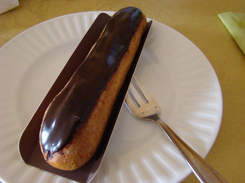
Holothuria edulis with the three players: the pink sausage, the gray one and the éclair. If one looks only at the ossicles, the character of choice to tell sea cucumber species apart, it seems like there is only one species. However, if one looks at the color of the animals and their ecology, it seems that the three players should be considered different species. Ideally, to distinguish species, it is good to have at least two independent characters that tell you the same story. Another important criterion to decide whether two individuals belong to the same species lies in whether they can produce fertile offspring. An efficient way to tell whether individuals can interbreed is to look at their DNA.


The gray form of Holothuria edulis. Cape Maeda, Okinawa, 15m. Photo by François Michonneau/FLMNH released under CC Atribution

How can a succession of A, T, C and G (the base pairs) help to distinguish between species? Just like you, each cell in the body of a sea cucumber contains DNA that it inherited from its parents when the sperm and the egg fused. To create a fully functional organism based on this single initial cell, the DNA has to be duplicated many times. Because each cell contains its own copy of DNA, the DNA is fully duplicated before each cell division. There are many mechanisms to ensure that the duplicated DNA is a perfect copy. However, on rare occasions, mistakes are made. DNA molecules are large and some parts are more important than others. In parts that are not very important, these mutations can accumulate without many consequences. In important parts, the slightest alteration can have important results. Because of these differences, not all parts of the DNA molecules evolve at the same speed. Some evolve so fast that they are unique to each individual and can be used in forensics to convict or acquit a suspect. Some change so little that they are almost identical across the entire tree of life.

Where mutations happen very rarely, if you find two individuals that share the same one in their DNA, they are more closely related than individuals that don't have this mistake. In other words, they share the same mistake because at some point in the past, their ancestors had the same parents. In animals cells, there is DNA in two compartments: the nucleus and the mitochondria. In the nucleus, each gene has two copies (one comes from mom, the other from dad) and the genes are arranged in long linear molecules: the chromosomes. The genes in the nucleus are responsible for most of the functions and appearance of the organism. Mitochondria are responsible for converting energy from sugars to make it available to the cells. In each mitochondrion, DNA is stored in a small single circular molecule and only contains genes that are useful to the mitochondrion. Unlike DNA in the nucleus, genes in the mitochondria are found in single copies: mitochondrial DNA from the parents don't mix, and only the mother contributes. These characteristics make the evolution of mitochondrial DNA easier to track and understand than nuclear DNA.

A typical animal cell showing the location of the nucleus and a mitochondrion. From NCBI.

Within the mitochondrial DNA, a gene has been widely used to help telling species apart. In most organisms, this gene accumulates mutations just at the right speed so that each species has a unique sequence. Because of this feature, the sequence of this gene can be considered as a "barcode". Just like each product at the supermarket has a unique sequence of numbers represented as a barcode, the sequence of this gene is unique to each species. However, contrary to the barcode that is found on all the packs of your favorite cookies, the barcode found in the mitochondrial DNA of a species is not perfectly identical from one individual to the next. Instead, out of the about 700 letters that make up the barcode, it is common to find a dozen of differences between the two sequences, but it's rare to find sequences that have more than 35 differences between two individuals of the same species.

Coming back to our species complex, what does the barcoding gene sequences have to say? The two most divergent barcode sequences that we have for the Holothuria edulis complex have about 20 differences, and they both belong to the pink sausage group. What is more surprising, is that the gray ones have exactly the same barcode sequence as some of the pink ones. The éclairs have their own unique sequence. However, they only have about 10 differences with some of the sequences from the pink ones.

If the barcode sequence was a perfect way to tell species apart, it would mean that the three color forms within this species complex are actually all the same species. However, it is not perfect. The three forms could actually be three good biological species that don't interbreed, and yet, their barcode sequence could say otherwise.

The first explanation to this pattern is these three form became different species very recently. Even when a species doesn't interbreed with another, it takes many generations for the barcode sequence to be completely unique and characteristic of this species. At first, when a pool of individuals start to diverge from the rest of the population, they will carry with them only a small sample of the barcode sequences that were characteristic of the ancestral population. Generation after generation, some of these sequences will go extinct (because the individuals carrying them didn't leave any descendants), and others will slowly accumulate mutations. Because these individuals don't interbreed with the rest of the population, these mutations will become characteristic of this new divergent species.

An alternative explanation could be that these three forms are actually different species but recently they swapped their mitochondrial DNA. In species that recently split, it can happen that the mechanisms preventing different species to interbreed fail. They can hybridize and in the process mix up their DNA, and in particular mitochondrial DNA. If it were the case, the signal shown by the barcode sequence could be misleading. The species may have stopped interbreeding a long time ago, but if they swapped their mitochondrial genes recently, the information from the barcode gene would be make us think that they belong to the same species.

The alternative to these hypotheses could be that the barcode gene is correct: the three forms are actually the same species and they just look different because they live in different habitats for instance.

Schematic reconstruction of what might be happening to the DNA of diverging populations. DNA is represented as a series of colored boxes. Each color represent one type of DNA base. A, B & C represent 3 lineages and each line correspond to a generation. At each generation an individual can leave one (or more) descendant with an identical copy of its DNA (white arrows), a descendant with a modified copy of its DNA (red arrow) or does not leave any descendants (crossed white arrows). At the fourth generation, lineages A & B cannot interbreed with C (represented by the dashed line). However, because of the recent history of their DNA, at the fourth and fifth generations B & C have more similar sequences than A. It would suggest, as in the first explanation, that B & C are more closely related despite the fact they cannot interbreed. Given enough time (generation 6 on the drawing) the lineages would reflect the correct relationships. If all the lineages could interbreed, DNA could still be exchanged. In this case B & C would seem closely related despite a history of reproductive isolation (as in the second explanation).

In the end, we are in a situation where color patterns and ecology say one thing (the three forms are different species) while ossicles and genetics suggest another (the three forms are the same species). Which is right? To understand what is happening in this particular case, in the next months, I am going to look at what nuclear genes have to say about it. Remember, mitochondrial genes only show a small part of the story as they are transferred only through the mothers. Nuclear genes, in particular those accumulating mutations faster than the barcode gene, could help explain what we observed in the mitochondrial DNA, and in turn, help us understand whether the pink sausage, the éclair and the gray one are the same species.

Sunday, May 29, 2011

Trip to Okinawa #1 -- How do you tell species apart?

I just got back from a trip to Okinawa. In the next week, I will report on the reason for my visit and some of the findings.

The island of Okinawa is part of the Ryukyu Islands, an archipelago in southern Japan.

For marine biologists, it is a very interesting place. First, the island is big enough to have lots of different types of habitats. Because many species are really picky about the place they call home, more kinds of habitats means more species are likely to be found. Second, Okinawa is at the northern limit of the Indo-West Pacific, the largest and most diverse marine biogeographic region.

The main goal of my visit was to collect specimens belonging to a species complex of sea cucumbers, and along the way compiling the list of all species of sea cucumbers that inhabit Okinawa.

The species complex I'm studying is called Holothuria edulis. In most areas of the Indo-Pacific, Holothuria edulis is easily identifiable. And for good reasons. It looks like a pink sausage that is burnt on one side. It prefers shallow places (less than 10 m / 30 ft) that are a little silty with few waves. That's why it is usually very common in lagoons and back-reefs. It's active both during the day and at night.

Holothuria edulis (the pink sausage). Photo by François Michonneau/FLMNH. CC.
In 2007, Mark O'Loughlin published with some other eminent sea cucumber biologists a new species that they named Holothuria nigralutea. The specimens were collected in Western Australia between 90 and 100 m (300 to 330 ft). At a first glance, it doesn't look very similar to the pink sausage Holothuria edulis. It is almost twice as long, it is light yellow with a series of black blotches on the dorsal side and a black stripe on the ventral side.

Holothuria nigralutea (the éclair). Dorsal view. Photo by François Michonneau/FLMNH. CC.

Holothuria nigralutea (the éclair). Ventral view. Photo by François Michonneau/FLMNH. CC.

To tell species apart, sea cucumber biologists like to look at ossicles. They are microscopic "bone"-like structures that are found in the skin of sea cucumbers. They can take all kinds of shapes and usually each species of sea cucumber is characterized by a unique set of ossicles. However, the ossicles of Holothuria nigralutea look very similar to the ones of Holothuria edulis.

Ossicles from Holothuria nigralutea (left) and Holothuria edulis (right). Fron O'Loughlin et al 2007.

So, last year while in Okinawa, we were very surprised and excited when one of our hosts, Daisuke Uyeno, brought us back 3 specimens of Holothuria nigralutea from his 45 m (145 ft) dive. Not only was it very far away from the location the species was first seen, but it was also much shallower. This considerably changed what we thought we knew about this species.

In Japan, biologists often give common names to species so that they can refer to them easily in Japanese. For Holothuria nigralutea the common name is "ekureanamako" or the éclair sea cucumber. Indeed, the black stripe on the ventral side of this species just looks like the delicious pastry.

Dark chocolate eclair from La Maison du Chocolat
An éclair by CraZeeCrafteeZ, on Flickr. CC.

After the pink sausage and the éclair, let me introduce you to the third player: the gray one. It likes the clear waters of the fore reefs exposed to waves. It is found usually below 10 m (30 ft) but not much deeper. It is a nocturnal beast. During the day it hides in crevices, but when the sun goes down it mops the reef frenetically (as much as a sea cucumber can be). It is also very easy to recognize: the dorsal side is taupe gray with white speckles while the ventral side is light beige. The gray one is found in some places of the Western Pacific: Micronesia, Cooks Islands, New Caledonia, Nauru and Okinawa. The gray one doesn't have a scientific name. Indeed, there is no physical difference other than the coloration that can tease this species apart from the pink Holothuria edulis.

The gray one. Photo by François Michonneau/FLMNH. CC.

To summarize we have three very different looking animals: the pink sausage, the éclair and the gray one. They are very different sizes: about 15 cm (6 in) for the pink one, 30 cm (12 in) for the gray one and 45 cm (18 in) for the éclair. They live in different places: shallow lagoons for the pink, exposed reef slopes for the gray one, and much deeper for the éclair. Yet, their anatomy is very similar and their ossicles are almost identical. So despite their apparent ecological differences, are they all the same species?

In the next post, we'll see what the DNA has to say about it.

Sources & Credits:
- P. Mark O’Loughlin, Gustav Paulay, Didier Vandenspiegel and Yves Samyn (2007). New Holothuria species from Australia (Echinodermata: Holothuroidea: Holothuriidae), with comments on the origin of deep and cool holothuriids. Memoirs of Museum Victoria. 64: 35-52. link | PDF
- All sea cucumber pictures by François Michonneau/FLMNH licensed under Creative Commons Attribution.
- Eclair picture by CraZeeCrafteeZ on Flickr licensed under Creative Commons BY-NC-ND.

Sunday, March 20, 2011

FUM 2 (FUM Rides Again)

This past February 19th our division hosted the second annual Florida United Malacologists (FUM) meeting over at Powell Hall. You might remember that several of our ranks attended last year's meeting in Sanibel (although I erroneously called it "Florida Union of Malacologists"). The first sign that something was up could be seen in the range.

That table is completely devoid of clutter! But it isn't the Twilight Zone, we tidied the place up. Another sign that we're hosting a meeting can be seen here:

Giving a presentation to a room full of malacologists, however friendly they might be, can be a nerve-wracking experience, and we have to keep our strength up with some fortifying snacks. The line of food actually extended on behind me, cutting the photo off at the [2 trays of] bagels was merely an aesthetic choice.

There were 15 presentations from various shelled-mollusk enthusiasts. Jodi and I actually gave our talks on slugs (no shells!) which were kindly received nonetheless. Here is John Starmer in action giving his talk on work that he and Chris Meyer did on Rapa snails.

That's right, Gustav's long-lost grad student John Starmer had returned to us just in time to be roped into a presentation, but he totally nailed it. In fact, all the presentations were interesting as demonstrated by this audience reaction shot.

We also had a good time socializing over lunch.

And dinner. (I know, I should really stop writing posts when I'm hungry).

Looking forward to seeing you all again at next year's meeting!

:) Mandy

Wednesday, March 16, 2011

Leg 2: deeper, colder, and with 80% more jellyfish

After a brief weather delay leg 2 commenced with 14 scientists on board and plans to explore the deeper northern waters of the Gulf. This included trawls up to 500 feet deep and dives up to 90 feet deep! We didn't know what to expect down there so François went into ninja mode.

Unfortunately for us, dive knives aren't an effective weapon against jellies. But once we made our way through smacks of them (that's right, a group of jellyfish is called a smack, I totally looked it up), the scenery on the bottom was actually quite nice.

We did a lot of dives at the Florida Middle Grounds where hard bottom substrate is covered with sponges, bryozoans, and soft corals. It was also really cold. Remember how I talked about leg one and how we were sooooo cold in 64 degree water? Well, this time my dive computer said 57. And my wetsuit wasn't any thicker. The cold is distracting, but so is all the cool stuff down there. For instance, check out this irregular urchin with his extra long spines.

And this anemone

Despite Rob's absence on this leg, we couldn't help but notice all the fish (even though we tried not to, they have a backbone and all).

Gustav also found a sea slug or two.

Back on the surface things were no less eventful. This is a picture of the frenzy on the deck after we return from a dive and everyone opens up their collecting bags to stabilize the animals that they have collected. The frenzy is also heightened by cold and usually meal anticipation.

Sand samples that we bring up are swirled in a giant bucket with water which is then sieved to extract animals that live in the sediment. Since polychaetes are commonly found using this method, Jenna is usually at the helm. In this instance she has recruited Antonio to help her.

After things are sorted on deck they are brought into the lab for processing. Although this picture makes it look fairly organized, don't be fooled. This picture captures an island of calm in the tempest of processing fever, which is also heightened by meal anticipation.

While in the lab one day busily anticipating the next meal we were also visited by a pod of dolphin. A few got pretty close to the boat.

All in all the trip was a great success and we found many animals that seem to be new records for the region; some might even be new to science. Thanks to all the visiting scientists for lending their expertise and thanks to the FIO crew for being the glue which holds the operation together, oil that keeps things running smoothly, and the hand that feeds us. For now, goodbye R/V Weatherbird II!

:) Mandy

Wednesday, March 9, 2011

They study what?

That's right, despite what you may have heard and believe, sponges are animals too! On this trip we were lucky enough to have three sponge experts on board. Brendan, Anna, and Nicole from FSU had the daunting task of processing and identifying the sponges we brought on board. Here they are staring at a dredge full of sponges and possibly wondering why Poseidon is punishing/blessing them with 10 gazillion sponges to process.

I realize that talk of sponges causes many people to react like this:

Even amongst invert lovers like us, sponges often remain a mystery, and I confess that while I might be able to slap a family or generic level identification on an arthropod or mollusc, the best I can do for sponges is "Porifera" (the phylum of all sponges, essentially identifying them as "sponge"). But both vertebrate and invertebrate marine biologists can agree that sponges are cool. Not only do they filter water in impressive volume and velocity (while still being sessile and lacking musclature to do so), in many of the areas we explored sponges provide most of the structure on the sea floor and are therefore important habitat. A single sponge can reveal a whole community of animals. Here is Rob slicing up a sponge looking for gobies (fish) which live within.

In addition to fish, sponges also host lots and lots of invertebrates, including this Synalpheus. If only Art had been there.

We also found this large and awesome nudibranch, Hypselodoris picta, which dines on sponges. Surpringly, even after seeing a sea slug this cool, Rob still insists on working on fish. I know, we couldn't believe it either.

Even trying to identify invertebrates from so many diverse phyla, many of the animals we study still have a body plan that is somewhat recognizable. They have characters based on feeding structures, tentacles, reproductive structures, color pattern, shape, claws. They often have something resembling a head or even a face. While color and shape play a role in sponge identification, most sponge characters are much more cryptic. Please forgive the blurriness of this picture, it was night and it was very choppy.

In addition to all the information we record about specimen location, habitat, depth, and so forth, the sponge team also records data on texture, compressibility, color, shape, and even smell. They take pictures underwater because many of these characters can change once the sponge is brought to the surface. When they get back to their lab at FSU they will also examine the microscopic spicules that many sponges have that are often an important character for sponge identification. These same spicules can also be a severe irritant often requiring sponge workers to wear gloves (and avoid rubbing their eyes, as I found out the hard way on a previous trip). While our work area was an explosion of marine life and field equipment that often spilled over into the two square feet of counter space that we generously allotted to Rob to work on fish, their work area looked like this, with many tiny cups filled with drying pieces of sponge:

Despite all these obstacles to studying sponges, look how happy Brendan and Anna are, possibly because they're about to go diving, possibly because no matter how many sponges they find on a dive they can't possibly bring back the work load that was pulled up in that trawl, and possibly because they know that when they get back it will be time for dinner!

No matter why they do it, we are grateful that they do. Not only are sponges important from a marine biologist's perspective, but their many toxic chemicals make them prime candidates for potential new medicines. Thanks sponge team!

:) Mandy

Saturday, March 5, 2011

Bon Voyage!

From March 4th through the 14th several representatives from the FLMNH, along with scientists from many other institutions, will be sailing the high seas on the R/V Weatherbird II of the Florida Institute of Oceanography. And by "high seas" I mean the Gulf of Mexico off the coast of Florida. We've been doing a lot of diving, but since I'm one of the divers (and therefore not wielding my camera) I hope you enjoy this picture of our dive gear.


We have had several dives a day for collecting. So far, they have usually been around 40-60 feet, with the temperature hovering around 64 degrees. Please take a minute to let that last number sink in. That's 64 degrees Fahrenheit. But while some of us are diving, others on the surface claim that they are actually too warm. I find that hard to believe, but Rob, the collections manager of the fish range at the museum is seen here wearing short sleeves while he tries to net a pelican.

Ok, so Rob is not actually trying to net the pelican, he's after floating clumps of sargassum and the fish hidden within. The pelican is probably after the same thing. In addition to the fish team composed of Rob and the pelican, we also have a sponge team, an amphipod soloist, a bryozoan soloist, and Nat, Jenna, Gustav, and I on general invert duty. There are 12 of us in total so that means a lot of specimens to sort and process back in the lab and a lot of data to keep track of.

So you'd better believe that at the end of the day we are ready for some hard-earned rest. Here's a picture of us at night.

I'm sorry, did I say rest? I meant trawl. The trawl was missing a part the first night, but we managed to get a few in tonight, and I have no doubt that we'll be making up for lost time. The trawl brings up a variety of fish as well as inverts, including the biggest sea star I have ever seen!

We've only just begun. I'll keep you posted!

:) Mandy